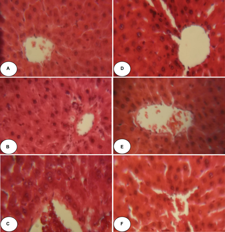
FIGURE 2.
Histopathological observation of rat hepatic tissue of control, ACEO, chlorpyrifos, and vitamin C stained with hematoxylin and eosin viewed at original magnification (×400). (A) Control liver showing normal architecture; (B) ACEO alone administered rats showing normal architecture in liver tissue; (C) CPF liver sections showed abnormal cellular morphology accompanied by sinusoidal dilatation, congestion, ballooning, necrosis, vacuole formations, and degeneration of hepatocytes after chlorpyrifos treatment; (D) ACEO + CPF co-treated rats indicating protected hepatic tissue; (E) VitC alone treated rats showed normal architecture in liver tissue; and (F) VitC + CPF showed that vitamin C co-administration restored liver tissue alteration induced by chlorpyrifos.

