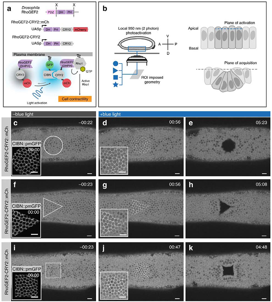
Figure 3. Optogenetics control over tissue invagination in developing drosophila.

Reproduced from Izquierdo et al., [43]. (A) Cartoon of genetically encodable system for optogenetic Rho1 activation. Cry2 of the blue light inducible dimerizing pair Cry2-CIBN is fused to DHPH catalytic domain of RhoGEF2, while CIBN is tethered to the plasma membrane. Two photon stimulation causes a change in Cry2 conformation, allowing it to bind CIBN, leading to recruitment of DHPH to the plasma membrane, where it can activate Rho1. (B) Cartoon showing experimental set-up. User-defined stimulation patterns can be applied to an epithelial sheet, triggering apical contraction and bending of stimulated cells out of the confocal acquisition plane. (C-K) Confocal images of RhoGEF2-Cry2-mCherry fluorescence integrated over 3 μM at the surface of the embryo. (C) Prior to photoactivation, the entire surface of the epithelial sheet is within the acquisition plane. (D) Upon photoactivation, RhoGEF2-Cry2-mCherry is enriched at the plasma membrane of cells within the circular activated region. (E) After sustained photoactivation for ~5 minutes, folding in the epithelial sheet caused by apical contraction in activated cells displaces the photoactivated region from the confocal plane of acquisition. Triangular (F-H) and square (I-K) geometries of photoactivation were also performed to demonstrate that arbitrary shapes of epithelial invagination could be specified by the user. Scale bars are 10 μM.
