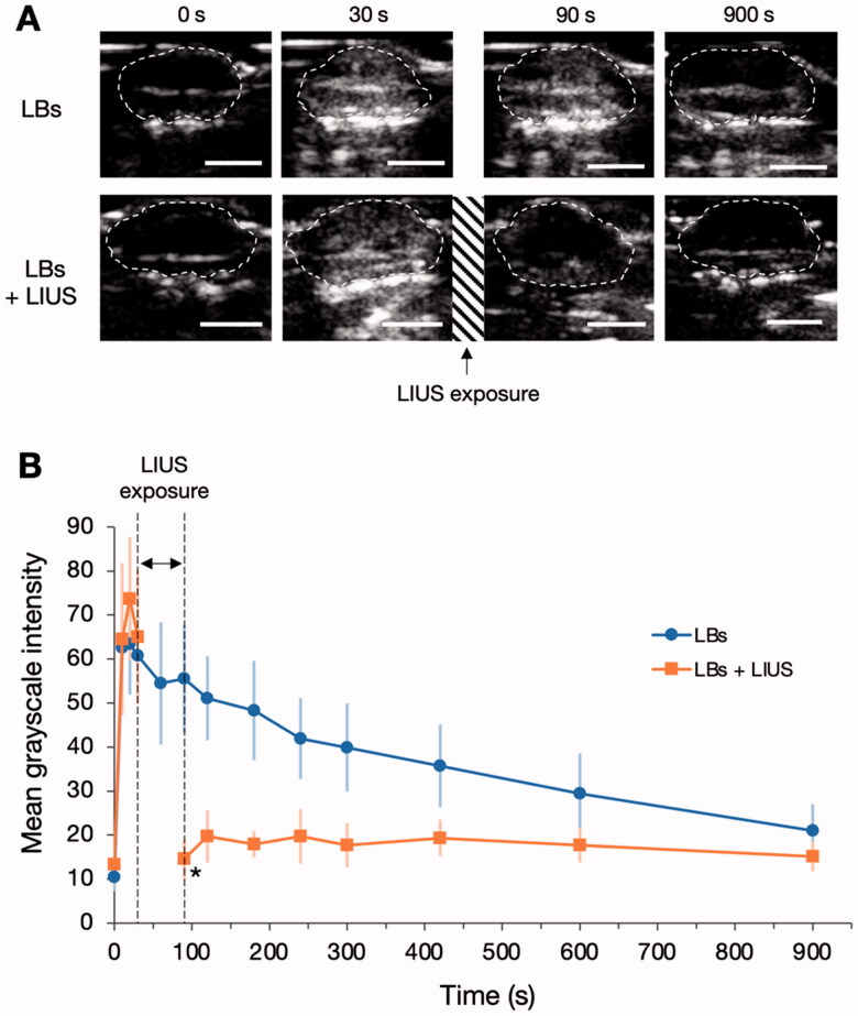
Figure 1.
Influence of low-intensity ultrasound (LIUS) on the stability of lipid bubbles (LBs) in the tumor tissue. (A) LBs were administered to mice through the tail vein, and contrast-enhanced images of the tumor tissues were visualized using an ultrasound imaging device for 900 s. The areas within the dashed lines indicate the tumor tissues. In the LBs + LIUS group, contrast-enhanced imaging was suspended between 30 and 90 s, and the tumor tissue was exposed to LIUS during that period. Scale bar: 5 mm. (B) Time-mean grayscale intensity (MGI) curves of the whole-tumor tissues in contrast-enhanced ultrasound imaging. MGI indicates the average degree of white signal in the tumor tissues and was calculated within the range of 0–255. Data are shown as mean ± standard deviation (n = 6). *p < .001 for the comparison between LBs and LBs + LIUs at 90 s, using Student’s t-test.

