Figure 1.
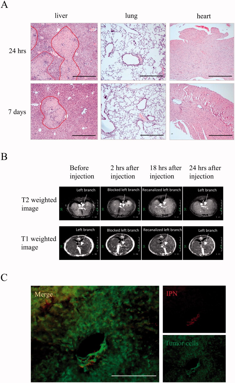
Severe hepatic necrosis in the left lobe of liver was induced by IPN hydrogel injection in early phase. (A) The hepatic histology was detected by H&E staining. Circled areas indicate hepatic necrosis. Bar = 400 μm. (B) The portal vein anatomy was scanned by MRI before and after (at 2, 18, and 24 hours) IPN hydrogel treatment. The upper panel was T2 weighted MRI images and the lower panel was T1 weighted MRI images. (C) The RFP-labeled hydrogels and GFP-labeled tumor cells in the tumor area at 2 weeks after IPN hydrogel treatment were detected by fluorescence microscope. Bar = 200 μm.

