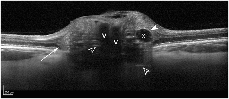
Figure 3.

Optic disc drusen morphology in a 22-year-old woman, shown with enhanced depth imaging optical coherence tomography. The drusen are seen as signal-poor structures (asterisk) with a partial hyper-reflective margin (solid white arrowhead). Prelaminar hyper-reflective lines (open white arrowheads) are thought to represent drusen precursors. Peripapillary hyper-reflective ovoid mass-like structures (longer white arrow) circumscribe the disc and likely represent herniated nerve fibres due to crowded optic nerve head conditions. Blood vessel shadows are denoted with v’s
