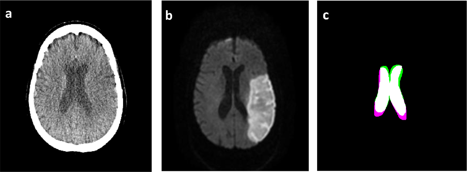
Figure 4:

Represents a slice from a CTP scan (a) following registration with a DWI volume whose corresponding slice is indicated in (b). Image (c) shows the spatial overlap of the ventricles from the two images with the CTP ventricles indicates in green, the DWI ventricle in pink, and the overlap of the two in white.
