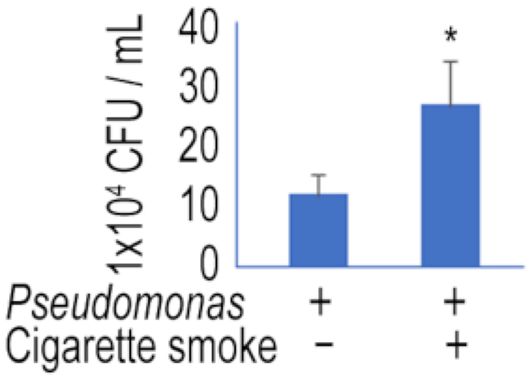
Figure 2: Drop plate method to determine bacterial load in lung epithelial BEAS-2B cells.

BEAS-2B cells were treated with 4% CSE for 3 h. Cells were then subjected to P. aeruginosa (strain PAO1) infection for 1 h followed by gentamycin treatment for another 1 h. Cells were lysed and the cell lysates were diluted to inoculate TSB plates for 16 h. Colonies were counted; the CFU numbers are illustrated in the plot. Graph shows mean ± SD, and “*” denotes P < 0.05. Results are representative of n = 3 experiments. Two-way unpaired Student t-test was used for smoke-treated and untreated groups. P < 0.05 indicates statistical significance.
