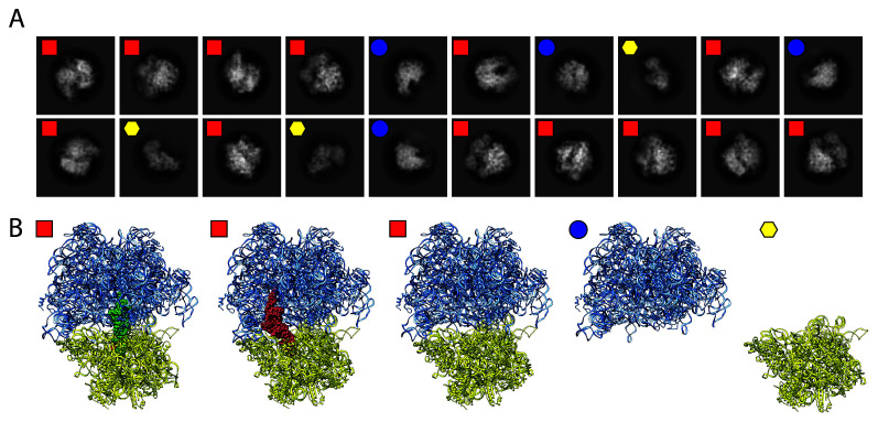
Figure 3. Ribosomal structures determined by single particle cryo-EM.
(A) Two-dimensional class averages obtained from a single cryo-EM experiment. Ribosomes were isolated from the Acinetobacter baumannii bacterium and flash-frozen onto a cryogenic grid. Shown are three separate classes that differentiate the 70S complex (red squares) from the individual 50S (blue circles) and the 30S (yellow hexagons) subunits. (B) Further computational sorting and analysis revealed three separate states of the intact 70S ribosome: tRNA bound at the P-site (green spheres), the E-site (red spheres), or empty. cryo-EM, cryo-electron microscopy.

