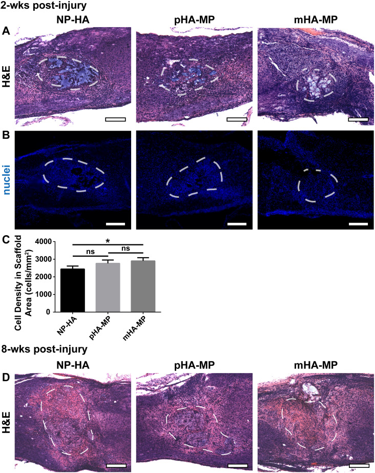
FIG. 4.
H&E and nuclei staining of spinal cord 2 and 8 weeks post-injury. Scaffolds were clearly identifiable 2 weeks post-injury in H&E stained sections, outlined in white dashed lines (a). Cells infiltrate into each scaffold type, though regions devoid of nuclei were observed, particularly in NP-HA scaffolds (b). Quantification of numbers of nuclei in scaffolds showed that mHA-MP scaffolds had significantly more cell infiltration than NP-HA, but not pHA-MP, scaffolds (c). Scaffolds were still identifiable 8 weeks post-injury in H&E stained sections (d) (*p < 0.05, Kruskal-Wallis test with Dunn's multiple comparisons test, n = 4–5, scale bars = 200 μm).

