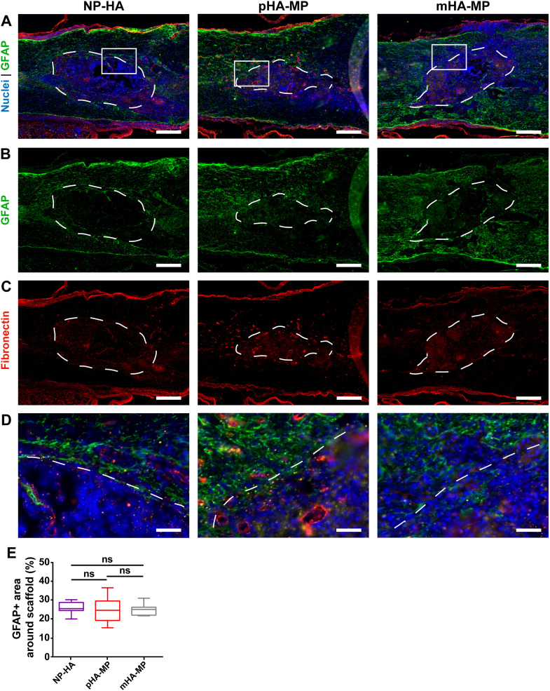
FIG. 6.
Immunostaining of astrocytic cell types and glial scar in scaffolds 2 weeks post-injury. GFAP+ astrocytes are found throughout the spinal cord (a), but denser staining was observed surrounding scaffolds (b), also associated with deposited fibronectin (c), consistent with previous work showing an activated astrocyte barrier. Zoomed insets (d) show scaffolds' borders. No significant differences in the GFAP+ area within 200 μm of scaffold borders were observed along scaffold types (e) [*p < 0.05, Kruskal-Wallis test with Dunn's multiple comparisons test, n = 5–6, scale bars = 200 μm (a)–(c), 40 μm (d)].

