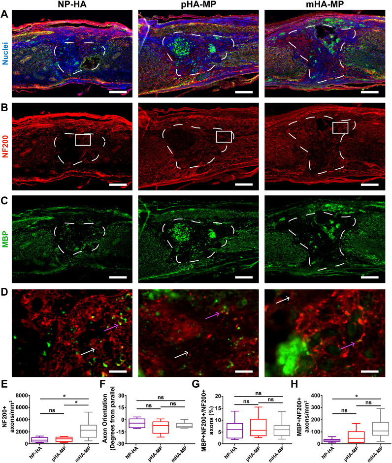
FIG. 7.
Immunostaining of myelinated axons in scaffolds 8 weeks post-injury. Spinal cords injected with NP-HA, pHA-MP, and mHA-MP scaffolds (a) showed clear scaffold boundaries that NF200+ axons (b) and MBP+ oligodendrocytes (c) appeared to cross. Zoomed-in images of the areas indicated by white boxes in (b) are shown in (d). White arrows indicate NF200+/MBP− axons, while pink arrows indicate NF200+/MBP+ axons. NF200+ axons had significantly greater densities in mHA-MP scaffolds than NP-HA or pHA-MP (e). The net orientation of axons was near parallel, relative to the longitudinal axis of the spinal cord, in each condition, with no significant differences among conditions (f). There was no difference in the percentage of axons that were myelinated (g). There was greater myelinated axon density in mHA-MP scaffolds than in NP-HA, but not pHA-MP, scaffolds (h) [*p < 0.05, Kruskal-Wallis test with Dunn's multiple comparisons test, n = 5–6, scale bars = 200 μm for (a)–(c) and 20 μm for (d), white arrows = unmyelinated axon and pink arrows = myelinated axon].

