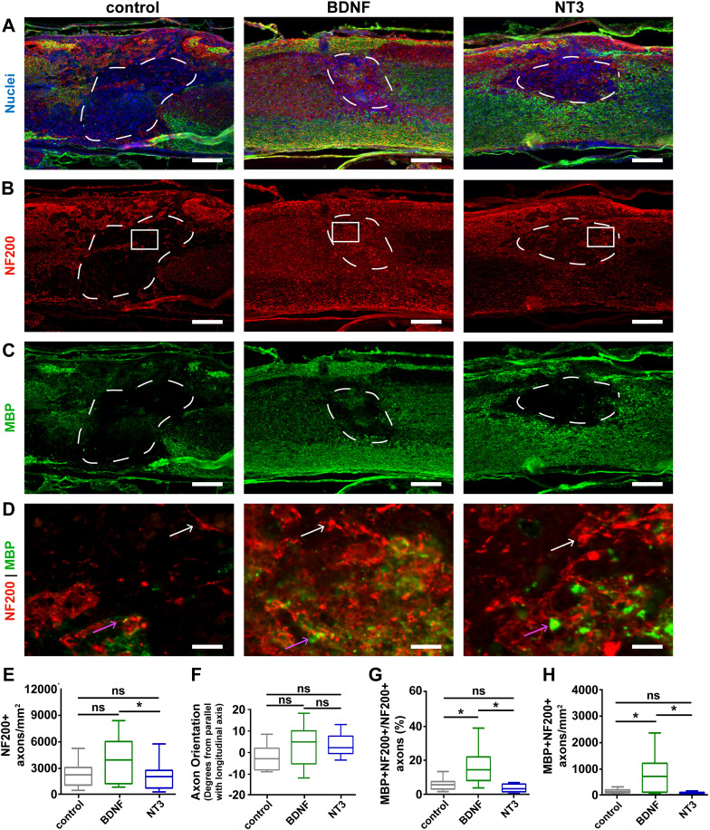
FIG. 8.
Quantification of myelinated axons after the delivery of neurotrophic factor-encoding vectors 8 weeks post-injury. mHA-MP scaffolds were delivered after injury containing BDNF, NT3, or control FLuc-encoding vectors (a) and showed NF200+ axons (b) and MBP+ oligodendrocytes (c) within the scaffolds. Zoomed-in images of the areas indicated by white boxes in (b) are shown in (d). White arrows indicate NF200+/MBP- axons, while pink arrows indicate NF200+/MBP+ axons. The delivery of vectors encoding for the BDNF increased the density of axons (NF200+) and myelinated axons (NF200+/MBP+). The axonal density was significantly higher with the delivery of the BDNF relative to NT3 (p < 0.1) but not FLuc control (e). The net orientation of axons was near parallel, relative to the longitudinal axis of the spinal cord, in each condition, with no significant differences among conditions (f). BDNF delivery led to increased proportion of myelinated axons (g) and densities of myelinated axons (h) relative to both NT3 and control ([p < 0.05, Kruskal-Wallis test with Dunn's multiple comparisons test, n = 5–6, scale bars = 200 μm for (a)–(c) and 20 μm for (d), white arrows = unmyelinated axon and pink arrows = myelinated axon].

