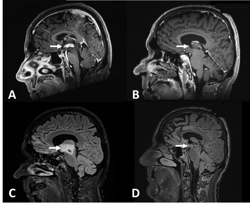
Figure 1. Sagittal MPRAGE MRI post administration of gadolinium contrast agent and T2 FLAIR sequences before and after the patient completed R-CHOP with dexamethasone treatment.
A: Sagittal magnetization-prepared rapid acquisition with gradient echo (MPRAGE) MRI shows presence of a large, contrast-enhancing third ventricular lesion. B. Two-week follow up MRI after completion of R-CHOP and steroid treatment, there is a significant reduction in lesion size. C/D: T2 fluid-attenuated inversion recovery (FLAIR) sequences show a similar degree of reduction in the volume of FLAIR hyperintensity.

