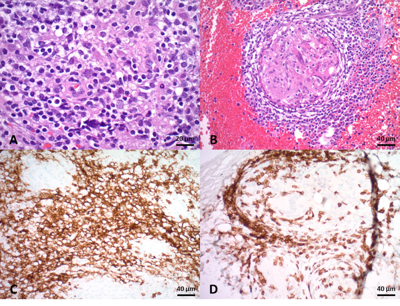
Figure 3. Histological analysis of the L5 lesion biopsy.
A: Sections showing patchy lymphomatous infiltration consisting of medium-to-large sized cells with scant eosinophilic cytoplasm and oval hyperchromatic nuclei with coarse chromatin with variable numbers of eosinophils. B: Scattered noncaseating granulomas are present in a background of lymphomatous infiltrate. C: Lymphocytic infiltrate stains positive for CD20. D: Lymphoid cells also stain positive for CD3 and surround a granuloma.

