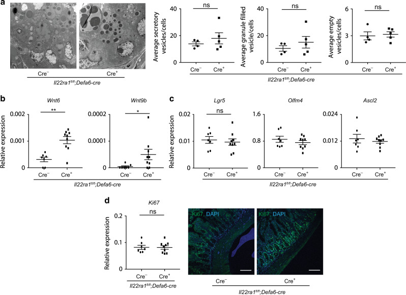
Fig. 5. Dysregulated expression of WNTs are present in Paneth cell-specific IL-22Ra1 knockout mice.
a Representative TEM images of Paneth cells (left panel) and number of secretory vesicles (right panel) in the terminal ileum of naive Il22Ra1fl/fl;Defa6-cre+/− mice. b RT-PCR analysis of Wnt6 and Wnt9b expression from terminal ileal tissues of naive Il22Ra1fl/fl;Defa6-cre+/− mice. c RT-PCR analysis of Lgr5, Olfm4 and Ascl2 expression from terminal ileal tissues of naive Il22Ra1fl/fl;Defa6-cre+/− mice. d RT-PCR and immunofluorescence analysis of Ki67 expression (left panel) and number of proliferative cells (right panel) from terminal ileal tissues of naive Il22Ra1fl/fl;Defa6-cre+/− mice. Figure 5b, c, d are generated from two independent experiments. Figure 5a is representative of at least 4–5 mice in each group. Data are presented as mean ± SEM on relevant graphs. Scale bars in relevant figures equal 100 μm. *P ≤ 0.05; **P ≤ 0.01 (Mann–Whitney test, two-tailed).

