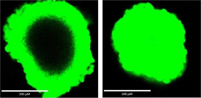
Figure 3.
Comparison of the ability of two different ATP assay reagents to lyse cells in ~ 350-μm diameter spheroids measured using staining with a vital fluorescent DNA binding dye. HCT116 cells were added to a hanging drop device (GravityPLUS™ 3D cell culture system from InSphero) and cultured for 4 d to produce ~ 350-μm diameter spheroids. The ATP assay reagents and nonpermeable DNA dye were combined and added according to the manufacturer’s recommended methods. The samples were shaken for 5 min followed by 25 min incubation before laser confocal microscopy was used to record photographs of the two spheroids using the same conditions.

