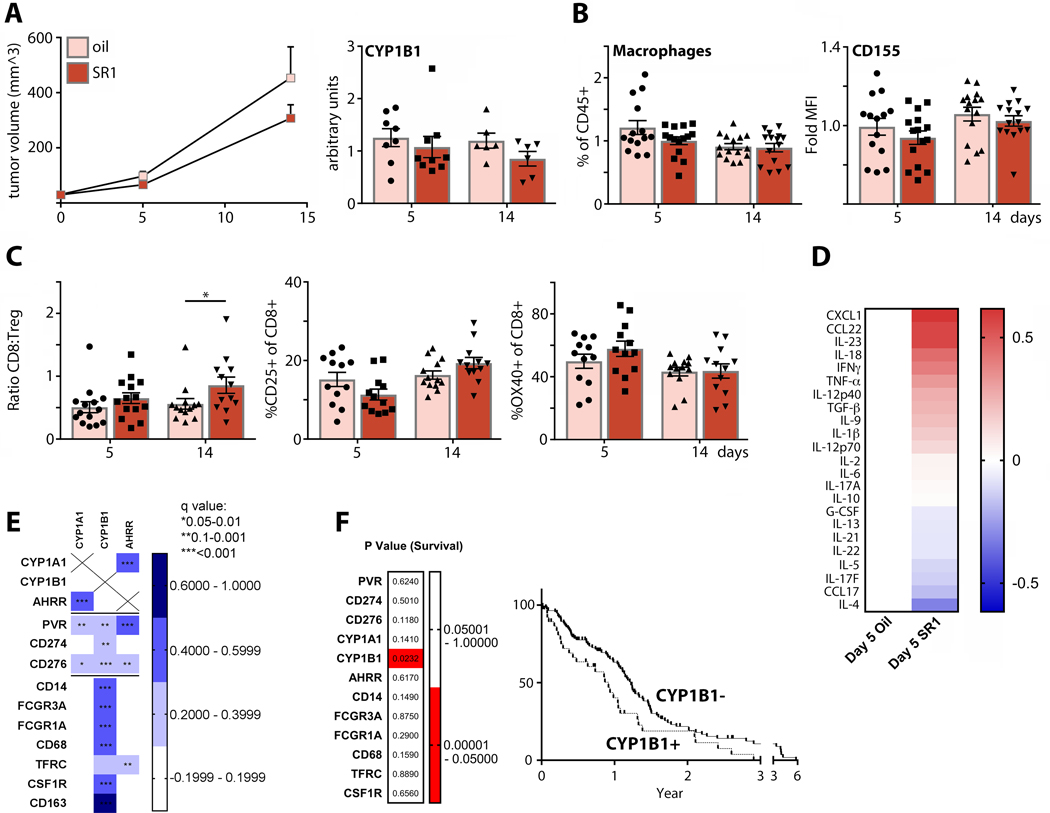
FIGURE 5.
AhR activity mediates tumor immunosuppression, and is associated with transcriptomic markers of TAMs and survival in GBM patients. (A; left panel) Mice bearing orthotopic E0771 tumors were treated every 48h with SR1 or vehicle control, and tumor volume was monitored (n=30 for day 5, n=15 for day 14). (Right panel) RT-qPCR was performed on tumor total RNA to assess CYP1B1 expression (n=8 for day 5 oil, n=9 for day 5 SR1, n=6 for day 14oil and SR1). (B) TAM (n=14 for day 5 oil, n=15 for day 5 SR1 and day 14 oil and SR1) and (C) intratumor T cell abundance (n=14 for day 5 oil and SR1, n=13 for day 14 oil, n=12 for day 14 SR1) were measured by flow cytometry. Tumor infiltrating T cell phenotyping was performed to assess changes in the CD8:TReg ratio, levels of CD25 expression, and OX40 expression. (D) Cytokine expression in post-dissociation tumor supernatant was measured by multiplex analysis and the ratio of vehicle (n=10) vs. SR1-treated (n=11) tumors plotted. (E) TCGA GBM database was analyzed for correlation of immune checkpoints (PVR, CD274, CD276), AhR activity markers (CYP1A1, CYP1B1, and AHRR), and macrophage markers (CD14, FCGR3A, FCGR1A, CD68, TFRC, CSF1R, and CD163). (F) TCGA GBM samples were separated by high (z-scores>0) and low (z-scores<0) expression of genes listed in (E) and compared for survival. Only sorting by high and low CYP1B1 expression yielded significant difference in survival.

