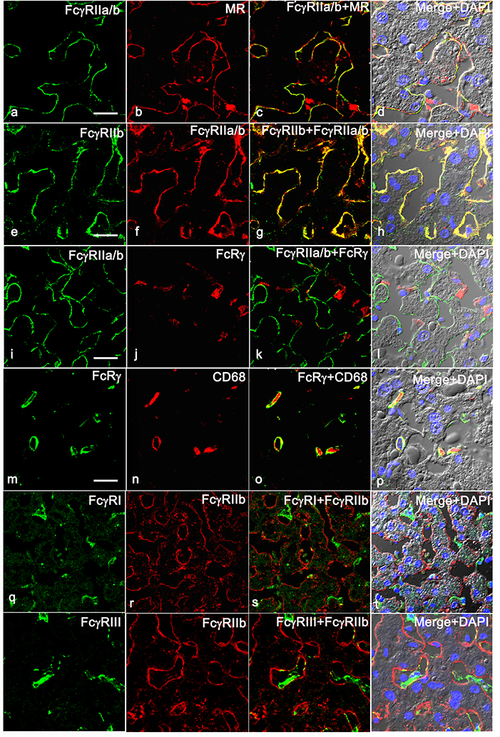
Figure 1. Human LSEC express only FcγRIIb.
Representative three-color immunofluorescence image from three biologically independent human liver samples. The 4 panel top row portrays the expression pattern of FcγRIIa/b in human liver using pan FcγRII mAb KB61 (green) along with LSEC marker anti-mannose receptor (MR) (red). The 2nd row shows the expression of FcγRIIb using pAb 163.96 (green) that colocalizes with mAb KB61 (red). The 3rd row shows the expression of FcγRIIa/b using mAb KB61 (green) along with anti- FcRγ (red). The 4th row illustrates the expression of FcRγ (green) along with KC marker CD68 (red). The 5th row illustrates the expression of FcγRI (green) along with FcγRIIb using pAb 163.96 (red). The 6th row demonstrates the expression of FcγRIII (green) along with FcγRIIb using pAb 163.96 (red). The column 3 shows the merged image of first two columns. Column 4 shows the merged images of the first two columns along with DIC and DAPI staining of nuclei (blue). The scale bar in 1st column indicates 20μm.

