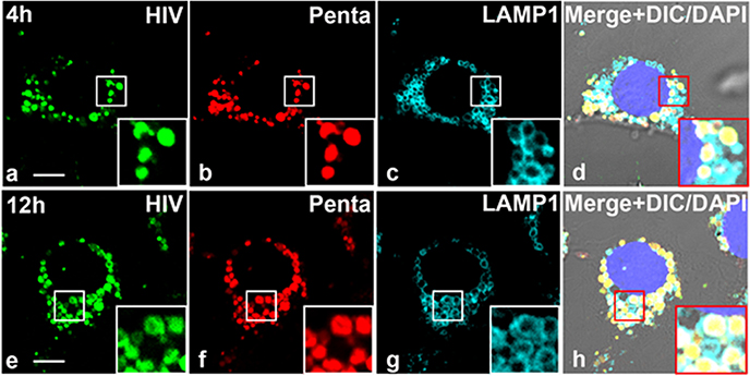
Figure 8. HIV-Pentamix immune complex localizes within lysosomes of LSEC.
A. Four-color fluorescence microscopic image of LSEC from 2B-KIX mice incubated with 488-HIV–647-Penta complex at 37°C and immunolabelled with anti-LAMP-1 antibody at 4hrs (Top Panel) and 12h (bottom panel). The data are representative of 15 images from two different mice/biological replicates. (a,e) Green puncta identify 488-HIV particles. (b,f) Pseudo colored Red puncta identifies 647-Penta mix. (c,g) Pseudo colored cyan identifies lysosome membrane structure marked by anti-LAMP-1 Ab. (d) Merged (a)–(c) plus DAPI stained nuclei and DIC. The scale bar represents 5 μm.

