
Figure 6. Intestinal lymph flow starts immediately after correct placement of catheter.
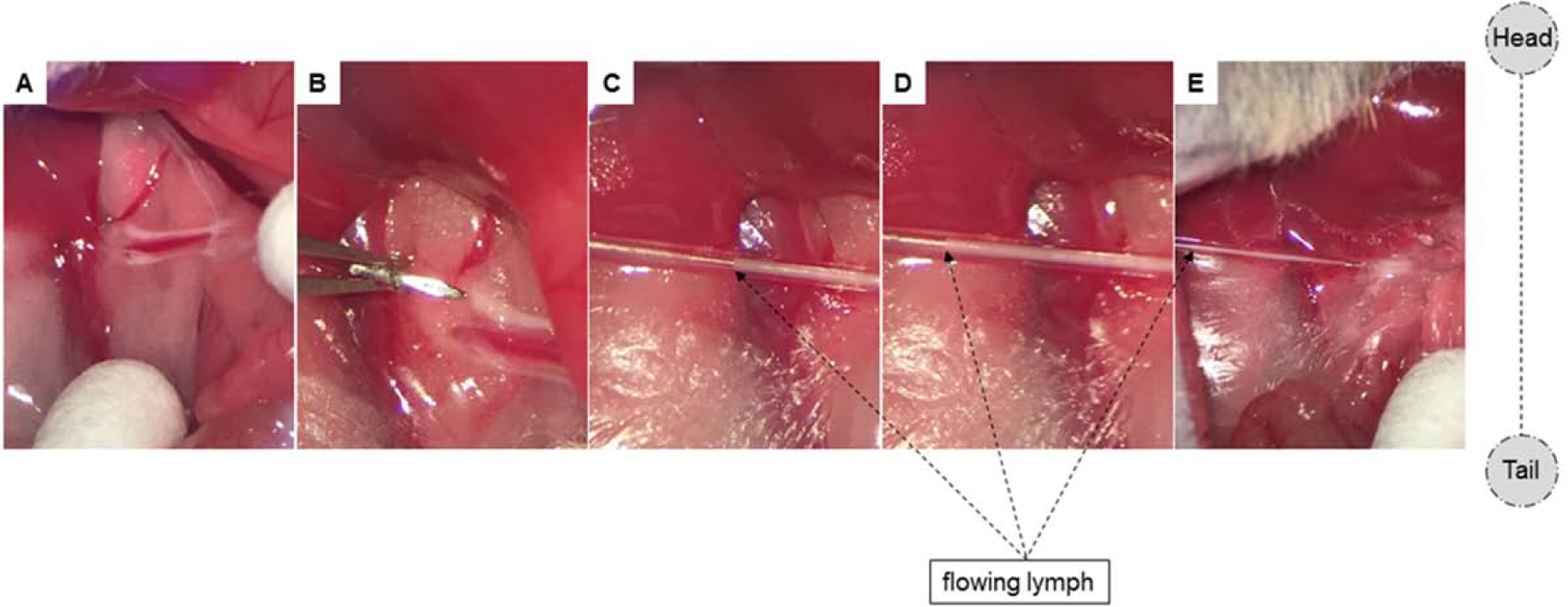
To augment the appearance of the lymph fluid for demonstrative purposes, commercially-available olive oil was administered 30min prior to cannulation. (A) Two MLDs are clearly visible with bright, white lymph fluid. (B-D) Immediately after lymphatic cannulation, lymph starts flowing. Pictures (C-E) were taken 3, 5, and 8 seconds after cannulation, respectively.