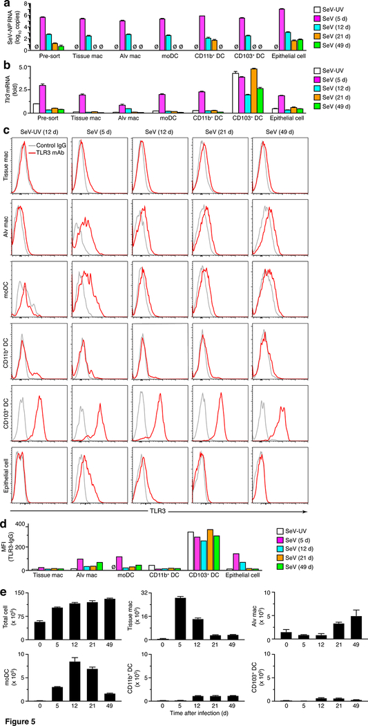
FIGURE 5.
Increases in viral RNA, TLR3 expression, and cell number implicate moDCs as an abundant cell site after viral infection. (a) Levels of SeV NP RNA in total cells and FACS-purified cell populations from lungs of WT mice at indicated times after SeV infection or SeV-UV. (b) Levels of Tlr3 mRNA for conditions in (a). (c) Levels of intracellular TLR3 for conditions in (a). (d) Quantification of MFI for TLR3 (expressed as the value for TLR3 minus IgG control) for conditions in (a). (e) Levels of total and immune cell populations in lung tissue from WT mice at 5–49 d after SeV infection or control SeV-UV (0 d). All data are representative of two to three separate experiments (mean and SEM) with at least 5 mice per condition in each experiment. *P<0.01.

