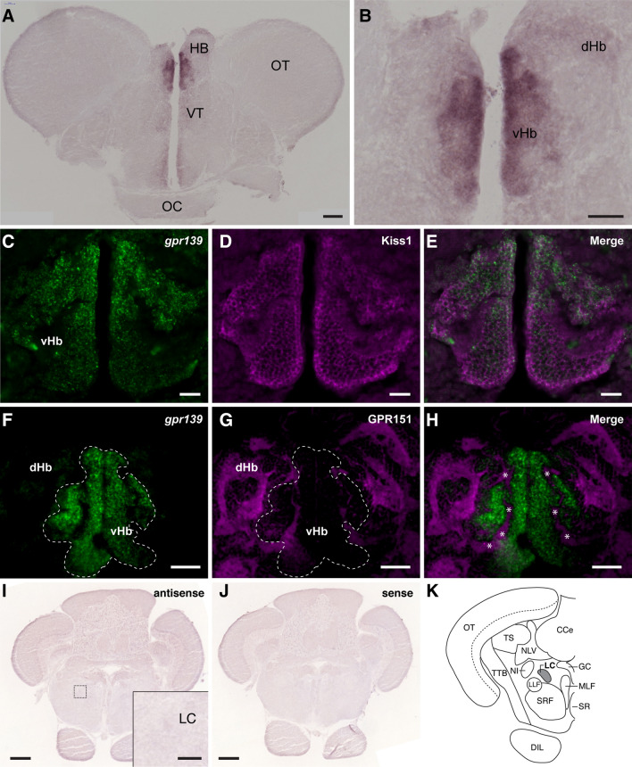
Figure 1.
Localisation of gpr139 mRNA expression in the brain of zebrafish. In situ hybridisation shows expression of gpr139 mRNA in the ventral habenula (vHb) (A, B). Double labelling of gpr139 mRNA (C, F) with Kiss1 (D) or GPR151(G) immunofluorescence further confirmed specific expression of gpr139 mRNA in the vHb (E) but not in the dorsal habenula (dHb) (H). In the vHb, GPR151-immunoreactive neuropil structure is also seen, which are derived from the dHb (H, asterisks). In the hindbrain region, no expression of gpr139 mRNA was detected in the locus coeruleus (I–K). The dotted box indicates the location for the inset in the panel I. HB habenula; VT ventral thalamus; OT optic tectum; OC optic chiasma; dHb dorsal habenula; vHb ventral habenula; LC locus coeruleus; TS torus semicircularis; CCe cerebellar corpus; NLV nucleus lateralis valvulae; GC central gray; MLF nucleus of the medial longitudinal fascicle; NI nucleus isthmi; LLF lateral longitudinal fascicle; SRF superior reticular formation; SR superior raphe nucleus; TTB tecto-bulbar tract; DIL, diffuse nucleus of inferior lobe. Scale bars: (A, F–H), 100 µm; (B) inset of (I), 50 µm; (C–E), 20 µm; (I, J), 200 µm.

