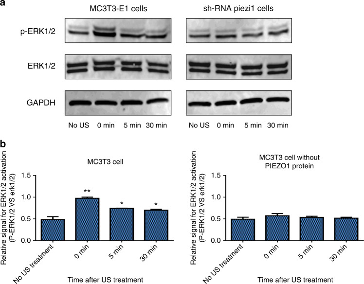
Fig. 4.
Western blot analysis of ERK1/2 and p-ERK1/2 in MC3T3-E1 and shRNA-Piezo1 cells after LIPUS stimulation. a Representative western blots of ERK1/2, p-ERK1/2, and GAPDH in MC3T3-E1 and shRNA-Piezo1 cells at the indicated time points (0, 5, and 30 min) after LIPUS stimulation. b Quantitative changes in ERK1/2 activation in MC3T3-E1 and shRNA-Piezo1 cells. The ratio of ERK1/2 phosphorylation to the relative expression of the protein doublet (p-ERK1/2 vs. ERK1/2) is presented as a parameter of ERK1/2 activation (n = 3, *P < 0.05, **P < 0.01, Student’s t-test)

