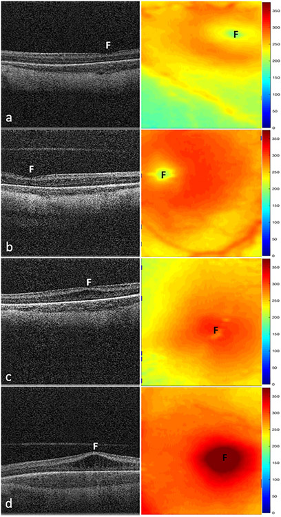
Figure 1.
Representative swept-source OCT B-scans (left column) and retinal thickness maps (right column) of macular edema severity in stage 2 ROP. A, Infant with no edema with a retinal thickness of 186 μm and inner nuclear layer (INL) thickness of 30 μm at the fovea (F). B, Infant with mild edema showing little to no deformation of the foveal contour with a retinal thickness of 212 μm and INL thickness of 52 μm. C, Infant with moderate edema showing flattening or slight upward bulging of the fovea and retina and INL thicknesses measuring 294 μm and 151 μm, respectively. D, Infant with severe edema showing severe upward bulging of the foveal contour with retinal and INL thicknesses of 433 μm and 286 μm, respectively. Mean retinal thickness at the fovea increased from 160 μm (standard deviation, 43 μm) without macular edema to 460 μm (standard deviation, 76 μm) with severe macular edema (P < 0.001).

