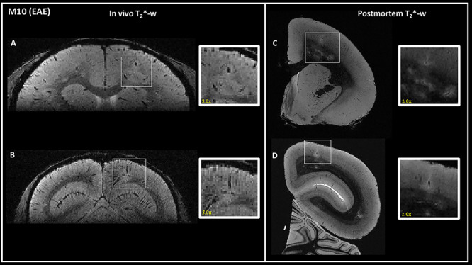
Figure 3.

Detection of the same cortical lesions in one EAE animal (M10) using in vivo (left) and postmortem (right) 3D T2*-weighted gradient-echo sequences. One intracortical (A) and one leukocortical (B) lesions were detected in vivo in two different areas: (A) right parietal cortex and (B) right occipital cortex. The same lesions were identified on postmortem MRI (C and D). In vivo lesions are magnified by a factor of three and postmortem lesions by a factor of two in the inset. Interestingly, cortical lesions appear larger in the in vivo images compared with postmortem images, likely due to water shifts.
