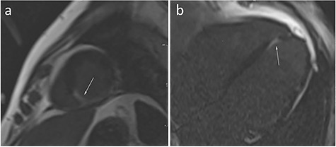
Figure 2.

Cardiac magnetic resonance imaging (CMRI). Late gadolinium enhancement images in the short axis (a), and four chamber (b) of the heart. The arrows point to the pathologic area of the myocardium (white area in the background of normal, black, myocardium).
