Fig. 2.
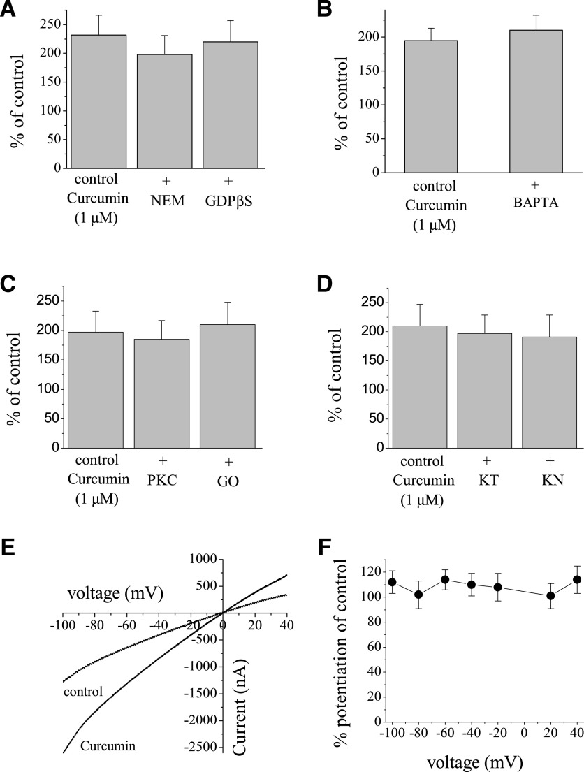
Effects of curcumin on α7-nACh receptor are not mediated by G-proteins and protein kinases, and are not dependent on intracellular Ca2+ levels and changes in membrane potential. (A) Bar presentation of the effects of 1 µM curcumin application (10 minutes) on the maximal amplitudes of ACh (100 µM)-induced currents in oocytes injected with 50 nl of distilled water, controls (n = 16), or NEM (10 mM, 50 nl, n = 8) and GDPβS (10 mM, 50 nl, n = 7) 30 minutes before recordings. (B) Bar presentation of the effects of 1 µM curcumin application (10 minutes) on the maximal amplitudes of ACh (100 µM)-induced currents in oocytes injected with 50 nl distilled water, controls (n = 5), or BAPTA (200 mM, 50 nl, n = 7). (C) Bar presentation of the effects of 1 µM curcumin on α7-nACh receptor–mediated currents in oocytes pretreated with vehicle (0.01% DMSO, n = 5) or PKC-412 (PKC; 10 µM, 30-minute pretreatment, n = 7), or Go-6983 (GO; 10 µM, 30-minute pretreatment, n = 6). (D) Bar presentation of the effects of 1 µM curcumin on α7-nACh receptor–mediated currents in oocytes pretreated with vehicle (0.01% DMSO, n = 7) or KT-5720 (KT; 10 µM, 30-minute pretreatment, n = 7) and KN-62 (KN; 50 µM, 30-minute pretreatment, n = 6). (E) Current-voltage relationships of acetylcholine-activated currents in the absence and presence of curcumin. Normalized currents activated by 30 μM ACh before (control, filled circles) and after 10-minute treatment with 1 μM curcumin (open circles). Each data point presents the normalized means and S.E.M. of seven experiments. (F) Quantitative presentation of the effect of curcumin as percentage of controls at different voltages.

