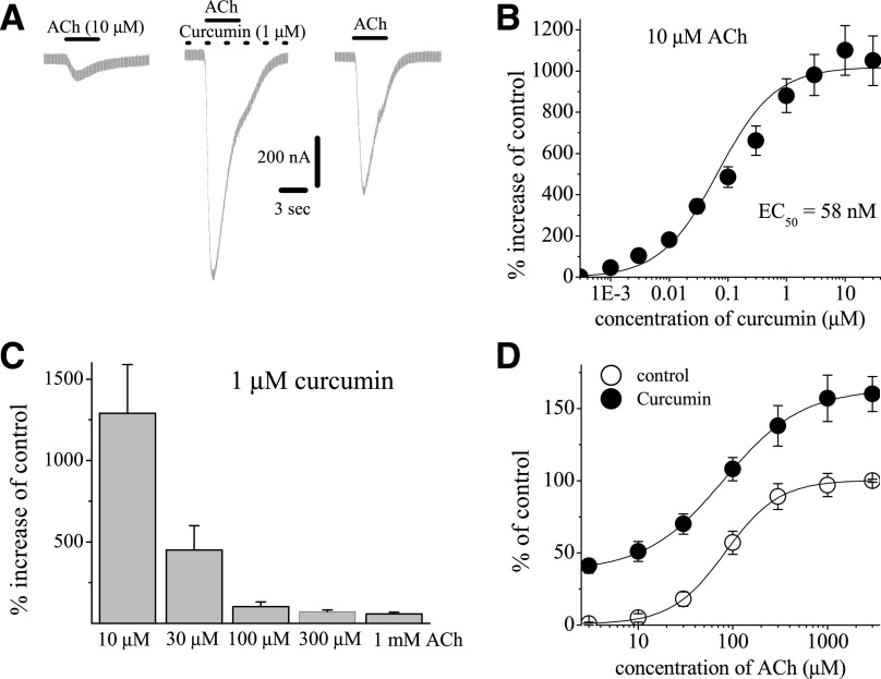
Fig. 3.
Effects of curcumin at different concentrations of acetylcholine. (A) Records of currents activated by ACh (10 μM) in control conditions (left), after 10-minute pretreatment with curcumin (1 µM) and coapplication of 1 μM curcumin and ACh (middle), and 10-minute washout (right). (B) Concentration-dependent effect of curcumin on α7-nACh receptors activated by low acetylcholine concentration. Each data point represents the mean ± S.E.M. of six to eight oocytes. The curve is the best fit of the data to the logistic equation described in the Materials and Methods section. (C) Bar presentation of the effect of curcumin at different acetylcholine concentrations. Bars represent the means ± S.E.M. of five to eight experiments. (D) Effect of curcumin on the ACh concentration-response relationship. Oocytes were voltage clamped at −70 mV, and currents were activated by applying ACh (1 μM to 3 mM). Oocytes were exposed to 10 µM curcumin for 10 minutes, and ACh was reapplied. Paired concentration-response curves were constructed and responses normalized to maximal response under control conditions. Data points obtained before (control) and after 10-minute treatment with curcumin (10 μM) are indicated by filled and open circles, respectively. Each data point presents the normalized means and S.E.M. of 7–11 experiments.

