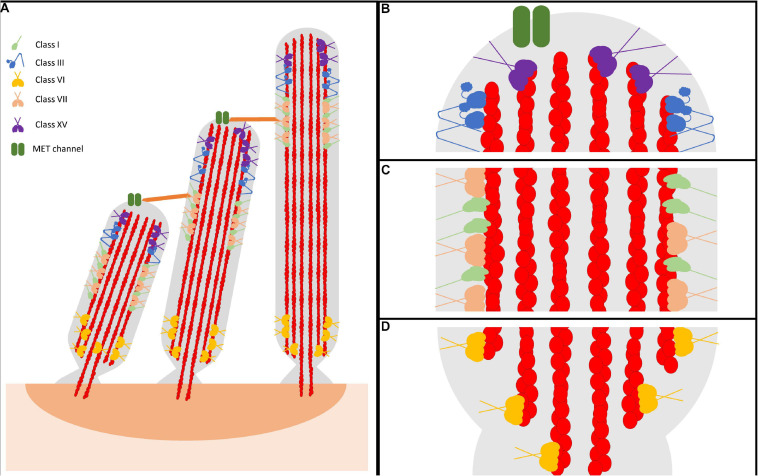
FIGURE 1.
Representative model of myosin motor localization in inner ear hair cell stereocilia. (A) Three rows of stereocilia form a staircase-like pattern, tethered together by various tip links, that is maintained through an organism’s lifetime. In an individual stereocilium, there are three main regions where myosins localize: (B) the stereocilia tips, also known as the lower tip link density, containing class III myosins and class XV myosins, as well as MET channels, (C) the lower tip link density, containing class I and class VII myosins, and (D) the anklet, containing class VI myosins. MYO7A has been shown to localize throughout the entire length of the stereocilia, however since its proposed function as a tensioning myosin occurs at the UTLD it is shown only here for simplicity. Apart from class III myosins, all stereocilia myosins have direct evidence of membrane binding. However, class III myosins are hypothesized to interact with the membrane, potentially modulated via its binding partner MORN4. Lastly, both MYO7A and MYO15A have not been directly shown to form dimers on their own, however MYO7A can form dimers via a cargo-mediated mechanism and MYO15A was proposed to oligmerize in a complex with cargo. For simplicity, both are represented as dimers.

