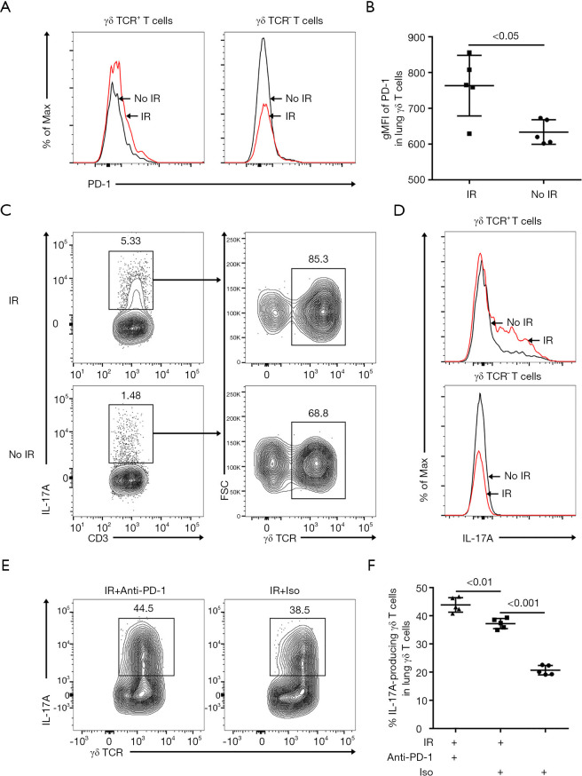
Figure 2.
Irradiation enhanced the expression of PD-1 on γδ T cells in murine lung tissues. (A) Representative histograms of PD-1 expression on CD3+ γδ TCR+ T cells or CD3+γδ TCR- T cells in lung tissues on day 7 post-irradiation. (B) gMFI of PD-1 were analyzed in lung γδ T cells on day 7 post-irradiation (n=5). (C) CD3+ IL-17A+ T cells were further divided into T cell subsets: CD3+ γδ TCR+ T cells and CD3+ γδ TCR- T cells. (D) Representative histograms displaying intracellular IL-17A staining of CD3+ γδ TCR+ T cells or CD3+ γδ TCR- T cells in lung tissues on day 7 post-irradiation. (E and F) The relative frequency of IL-17A-producing γδ T cells in lung γδ T cells on day 7 post-irradiation (n=5). PD-1, programmed death 1; gMFI, geometric mean fluorescence intensity; IL-17A, interleukin-17A.

