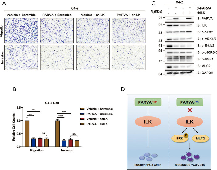
Figure 6.
ILK was essential for the metastasis-suppressing function of PARVA. Control or PARVA overexpression C4-2 cells were transfected with scramble or shILK#1. (A,B) Cell migration and invasion were analyzed by Transwell migration and Matrigel invasion assays. Representative images (A) and quantification (B) are shown. In each experiment, cells were counted in five random fields of each filter under a microscope using a 100× magnification. (C) Western blot analysis of ILK, p-c-Raf, p-MEK1/2, p-ERK1/2, ERK1/2, p90RSK, p-MSK1, MLC2, and PARVA expression. GAPDH was used as a loading control. (D) Schematic drawing depicting a model of PARVA-ILK-MAPK/ERK signaling axis in the regulation of PCa metastasis. PCa cells expressing a high level of PARVA, the binding of PARVA to ILK inhibited its downstream MAPK/ERK signaling and thereby attenuated metastasis. In a majority of PARVA-deficient PCa-tissues, lacking sufficient PARVA released the inhibition of ILK activity, thereby enhancing MAPK/ERK signaling and driving PCa metastasis. ***, P<0.001; ****, P<0.0001. ns, no significance; PCa, prostate cancer.

