Figure 6.
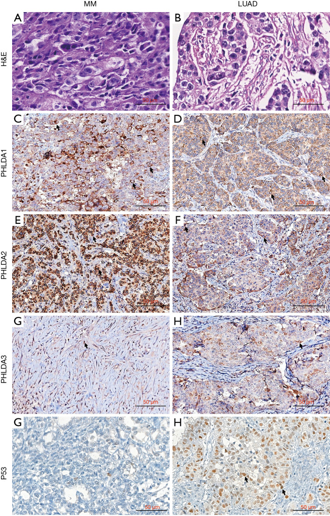
PHLDA protein expression using immunohistochemistry on tissue microarrays (TMAs) in MM and LUAD. H&E staining of an epithelioid MM section/showing blocks of epithelioid cells infiltrating sparse desmoplasic stroma (A); H&E staining of a representative solid subtype LUAD composed of cohesive and poor differentiated malignant cells, aggregated in blocks and immersed in exuberant desmoplasic stroma (B). Immunohistochemistry: PHLDA1 staining of mild number of epithelioid MM cells (arrows, C) and LUAD cells (arrows, D); PHLDA2 with strong and diffuse cytoplasmic staining with perinuclear accentuation in numerous MM malignant cells (arrows, E), contrasting with diffuse cytoplasmic staining in a mild number of LUAD malignant cells (arrows, F); PHLDA3 with diffuse cytoplasmic expression in MM malignant cells (arrows, G), and cytoplasmic staining with perinuclear accentuation in a mild number of LUAD malignant cells (arrows, H); strong and diffuse P53 nuclear staining in LUAD (arrows, J) and weak, almost absent, in MM (I). PHLDA, pleckstrin homology domain family A; H&E, hematoxylin & eosin; LUAD, lung adenocarcinoma; MM, malignant mesothelioma.

