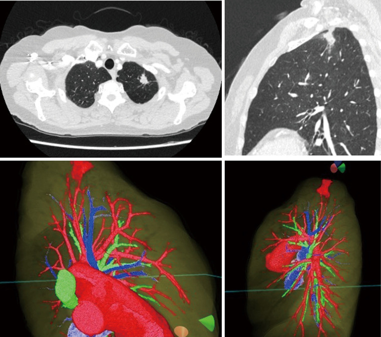
Figure 1.
A virtual dynamic image is generated from patient-specific 3D-CT data. The original preoperative chest CT scan demonstrating a left upper lobe tumor is shown (top row). Representative images of the 3D model (bottom row) demonstrate the anatomic relationship between the tumor and pulmonary anatomy.

