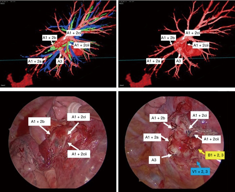
Figure 3.
Correlation of preoperative reconstruction and intraoperative findings. Representative images of the 3D model demonstrate high-resolution anatomical details of sub-subsegmental pulmonary artery branches (A1+2ci, A1+2cii) (top row). Intraoperative VATS images confirm the same vessel orientation as in the 3D model, prior to and after vessel ligation (bottom row).

