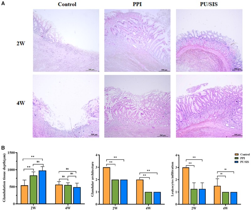
Figure 7.
Histological and morphological evaluation of gastric ulcer margin and ulcer base. (A) H&E staining of gastric ulcer on 2 W and 4 W post operation, respectively. Scale bars, 500 μm. (B) H&E staining and scoring in terms of the ulcer margin granulation tissue depth, and inflammatory infiltration. *: significantly different from control (P < 0.05). **: significantly different from control (P < 0.01).

