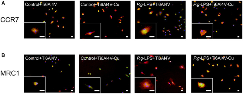
Figure 5.
Immunofluorescence staining of polarization-related markers in osteoporotic macrophages on Ti6Al4V and Ti6Al4V-Cu in response to P.g-LPS stimulation. Representative images of magnified views of macrophages are also shown. CCR7 (A) and MRC1 (B) were stained green by the use of Alexa Fluor 488-conjugated secondary antibodies, cytoskeletons were stained red with phalloidin and the nuclei were stained blue with DAPI. Bars = 2 μm

