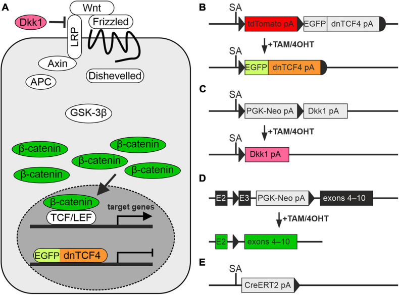
FIGURE 1.
Modulation of the Wnt signaling pathway in transgenic mice. (A) A simplified scheme of the canonical Wnt signaling pathway that shows by color-coding where the alterations in the cascade take place. (B–E) Schemes depicting genetic modifications present in the mouse strains used in the study. (B) The transcription of the Wnt-responsive genes in the nucleus is blocked by dominant negative T-Cell Factor 4 (dnTCF4; orange) fused to the enhanced green fluorescent protein (EGFP; light green). (C) Suppression of Wnt signaling at the membrane receptor level, due to the overexpression of a secreted Wnt inhibitor Dickkopf 1 (Dkk1; dark pink). (D) Activation of the pathway by the production of a stable variant of β-catenin. Cre-mediated excision of exon 3 (E3) removes the amino acid sequence involved in the degradation of the protein, and thus stable β-catenin aberrantly hyper-activates Wnt target genes (dark green). (E) Cre deletor mouse Rosa26-CreERT2 carries the gene encoding tamoxifen/4-hydroxytamoxifen (TAM/4OHT)-inducible Cre recombinase, fused with a mutated form of the estrogen receptor (CreERT2). APC, adenomatous polyposis coli; E2-3, exons 2-3; GSK-3β, glycogen synthase kinase 3β; LRP, low-density lipoprotein receptor-related protein; pA, polyadenylation site; PGK-Neo, neomycin resistance cassette; SA, splice acceptor site; TCF/LEF, T-Cell Factor/Lymphoid Enhancer Factor; td, tandem dimer; Wnt, Wingless/Integrated.

