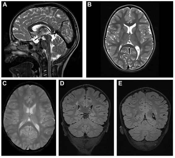
FIGURE 2.

Cerebral MRI. (A) T2-weighted sagittal TSE image of the patient shows a slightly thinned but fully formed corpus callosum and normal cerebellar structures. (B) T2-weighted axial TSE image of the patient reveals normal gyral pattern of the cerebral cortex and normal volume of white matter but delayed myelination of the internal capsules. (C) T2-weighted axial GRE image (T2∗) shows diffuse white matter abnormalities in the periventricular and subcortical. (D,E) T2-weighted coronal 3D-FLAIR images demonstrate focal as well as diffuse areas of hyperintensity in periventricular white matter.
