
Figure 4 (Top Panel):
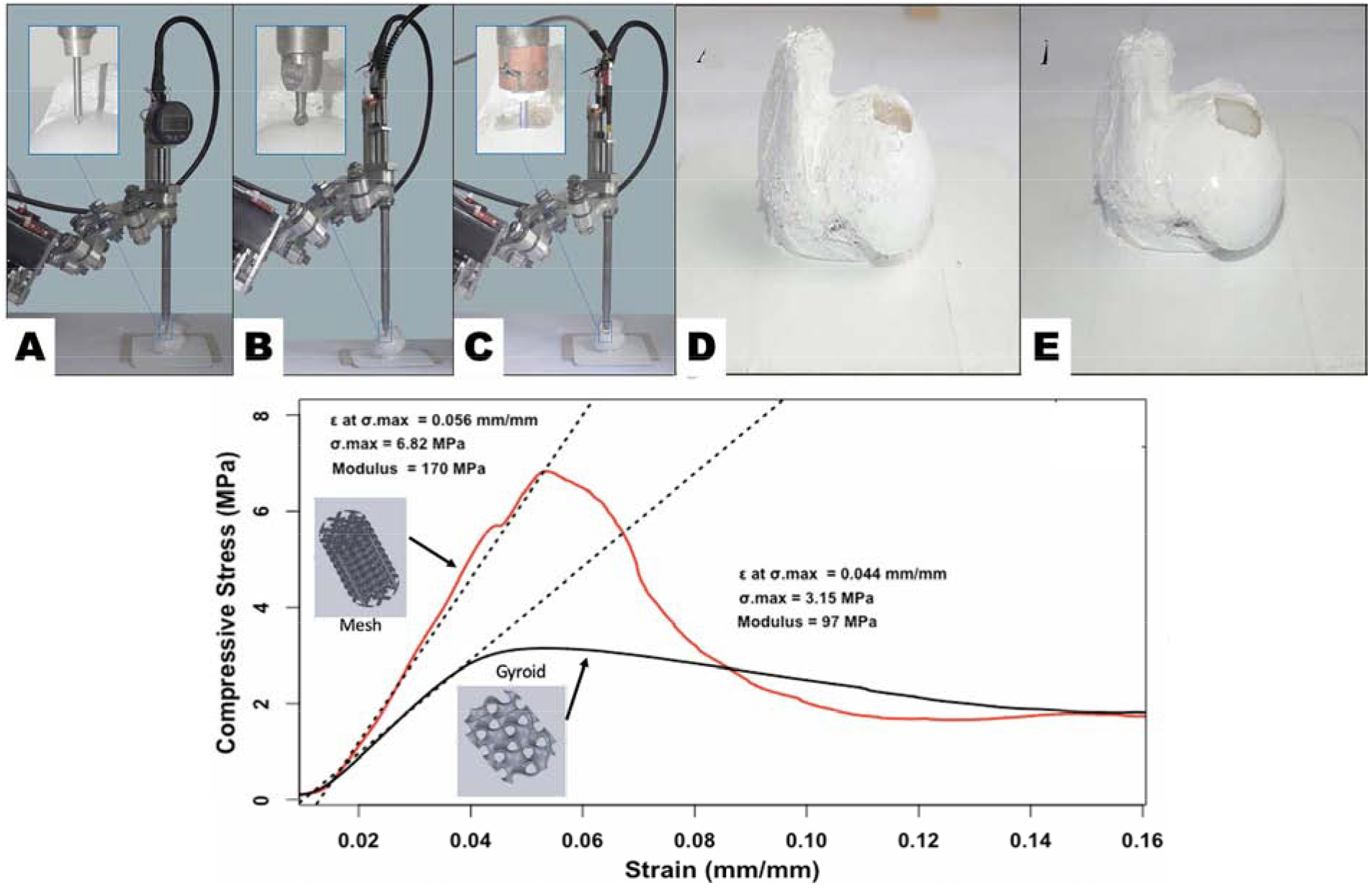
Experimental arrangement (A) Surface registration. (B) bone milling. (C) 3D printing. Bone samples pre and post 3D printing process (D) Milled defect sample. (E) Hydrogel infill sample. Both figures are reproduced with permission from [34]. (Bottom Panel): Compressive strength graph of two 3D printed pattern types based on data obtained from mechanical testing at UTEP conducted for reference.