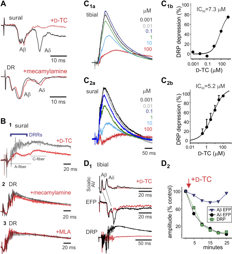
Figure 3.
The nicotinic acetylcholine receptor (nAChR) antagonist tubocurarine (d-TC) strongly depresses Aδ synaptic transmission and associated dorsal root potentials (DRPs). A: d-TC preferentially depressed the Aδ-fiber component of the extracellular field potentials (EFP), whereas mecamylamine did not alter EFPs. Stimuli are sural (4 T) and dorsal root (DR; 200 µA/500 µA). B: depression of evoked DRPs by nAChR antagonists. d-TC (1) and mecamylamine (2) depressed evoked DRPs, whereas methyllycaconitine (MLA) (3) was without effect (100 μA/100 μs, 10 T, and 100 μA/100 μs, respectively). Note that d-TC depression of DRP was associated with loss of dorsal root reflexes (DRRs). C: DRP was reduced by d-TC in a dose-dependent manner. Tibial (4 T) (C1) and sural (4 T) (C2) stimulation-evoked L3 DRP depression by d-TC. Dose-response curves for d-TC inhibition of tibial (C1b) and sural (C2b) nerve stimulation-evoked DRP. Tibial dose-response curve is from recording in C1a (12 episodes averaged). Sural dose-response curves are from four experiments (means ± SE), with example shown in C2a. D: relating depression of Aδ-mediated EFP to reductions in DRP amplitude. D1: d-TC depression of Aδ afferent EFP and DRP occur with similar magnitude and time course. Shown are the sciatic nerve afferent volley (AV), EFP, and DRP following stimulation of the tibial nerve at 4 T. Averaged responses are control and 20 min after bath application of d-TC. Note that sciatic nerve afferent fiber volleys are unchanged (without evoked DRRs), whereas L4 EFP and DRP depression occur. D2: amplitudes of evoked Aβ and Aδ EFPs and DRP are normalized to compare the relative time course and magnitude of depression produced by d-TC. Note that Aβ synaptic responses are relatively unchanged, whereas depression of Aδ EFP and DRP is closely matched. Data are from P6–8 mice.

