FIGURE 3.

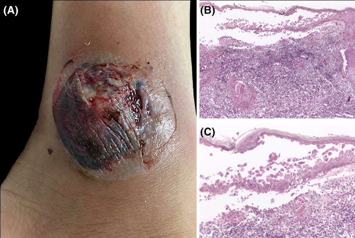
Bullous PG. A, Ankle with a tense hemorrhagic blister and adjacent ulceration with fibrin deposits. B, Biopsy shows intraepidermal bulla with acantholytic cells. C, Dense infiltrate of neutrophils with few eosinophils present in the dermis

Bullous PG. A, Ankle with a tense hemorrhagic blister and adjacent ulceration with fibrin deposits. B, Biopsy shows intraepidermal bulla with acantholytic cells. C, Dense infiltrate of neutrophils with few eosinophils present in the dermis