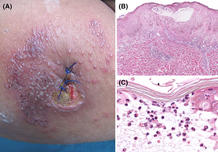
FIGURE 5.

Pustular PG. A, Leg ulcer with fibrinous wound bed and raised erythematous border. Numerous pustules surrounding the ulcer, which appeared just after the biopsy was taken. B, Low‐power view of a biospy showing hyperplastic epidermis with an intraepidermal pustule. C, High‐power shows the blister cavity containing numerous neutrophils
