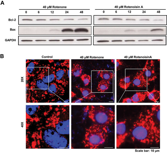
Fig. 4.

Rotenoisin A decreased Bcl-2/Bax and disrupted the mitochondrial network in MCF-7 cells. (A) Representative images of the western blot analysis. After rotenoisin A treatment, the expressions of Bcl-2 and Bax in MCF-7 cells shifted in a time-dependent manner. (B) Representative images of MitoTracker™ staining. The nuclei were counterstained with DAPI. Scale bar: 10 μm.
