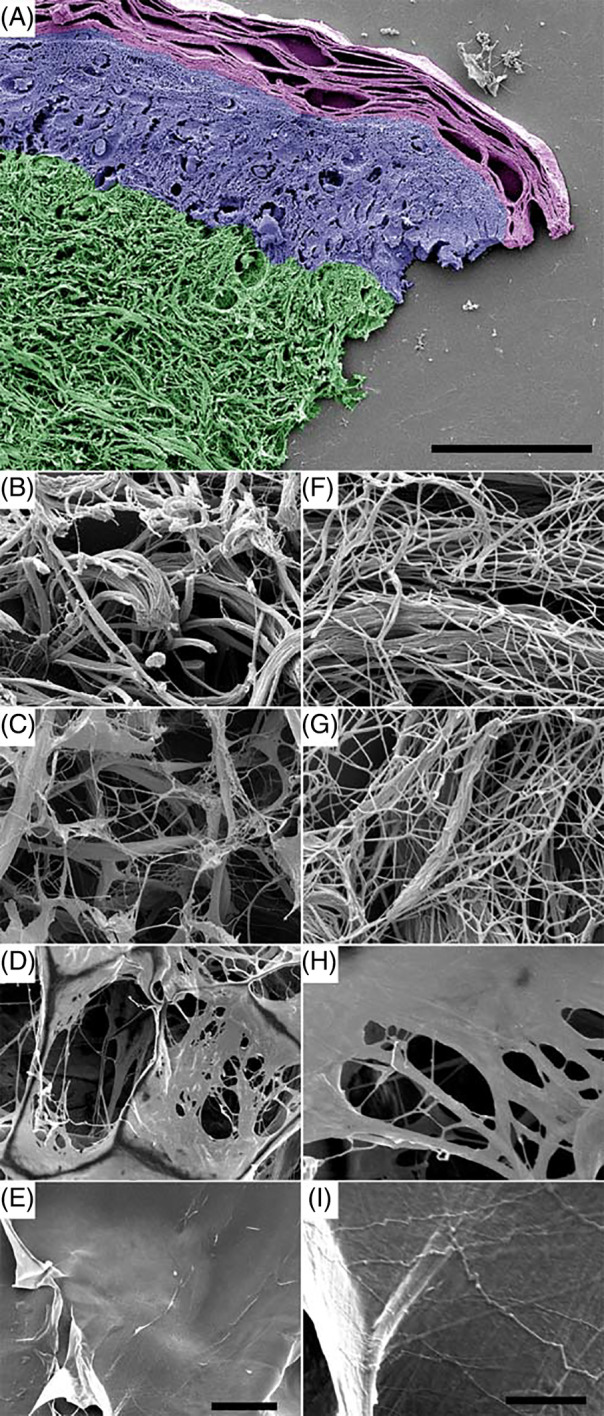
Figure 1.

Ultrastructure of different biomaterials as compared with native human dermis. Specimens of human skin (A, B, F) were prepared for field emission scanning electron microscopy (FESEM) and compared with collagen elastin matrix C‐E (C, G), bovine collagen matrix C‐GAG (D, H), and porcine collagen matrix C (E, I). In (A), the stratum corneum is represented in magenta, the epidermis in blue, and the dermis in green pseudocolour, respectively. Note the appearance of extended fibrillar collagen networks in the dermis (B, C) and in C‐E (F, G) and an abundance of amorphous sheets in C‐GAG (D, E) and C (H, I). Left lane, low magnification overviews (500×); right lane, high magnification of the same area (10 000×). The scale bars represent 50 μm (A), 20 μm (B‐E, left panel), and 5 μm (F‐I, right panel)
