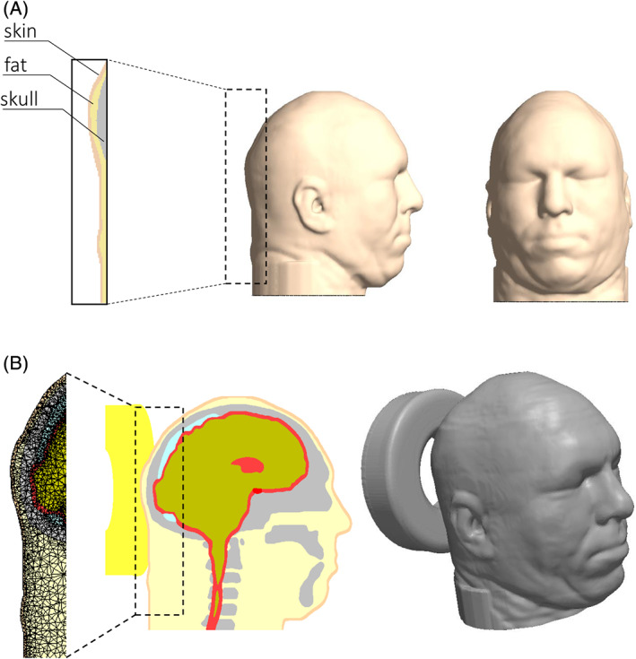
Figure 1.

Model geometry: A, side (left frame) and frontal (right frame) views of the head model, with magnification of the tissues at the back of the head (zoom‐in from the left frame). B, Mid‐sagittal cross section through the head model (left frame) and its three‐dimensional reconstruction when supported by the donut‐shaped gel head support (right frame); a magnification of the finite element mesh shows meshing at the occipital scalp region (zoom‐in from the left frame)
