Figure 2.

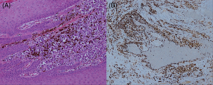
(A) Atypical melanocytes lining the dermoepidermal junction and extending into the dermis (haematoxylin eosin staining, ×100 magnification); (B) diffuse positivity in the tumour cells (HMB‐45 stain, ×40 magnification)

(A) Atypical melanocytes lining the dermoepidermal junction and extending into the dermis (haematoxylin eosin staining, ×100 magnification); (B) diffuse positivity in the tumour cells (HMB‐45 stain, ×40 magnification)