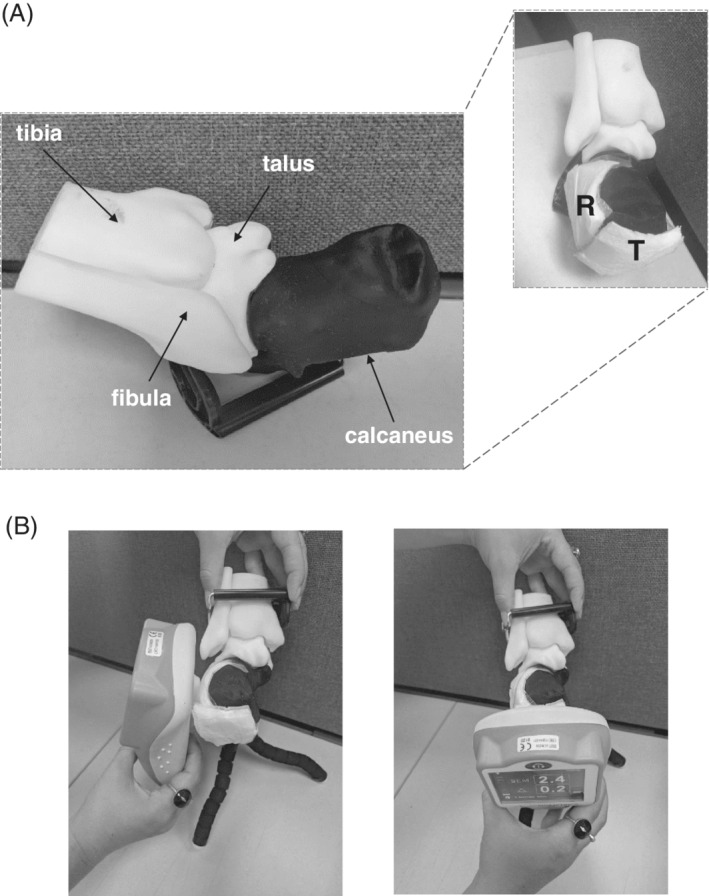
Figure 2.

The heel phantom model: A, A three‐dimensionally (3D) printed heel skeleton model (left frame). For the heel sub‐epidermal moisture (SEM) measurements, a reference diaper sample was placed at the lateral left aspect of the calcaneus and a test sample was attached at the posterior calcaneal aspect (right frame). B, Acquisition of SEM readings at the reference (left frame) and test (right frame) locations. T = test and R = reference samples
