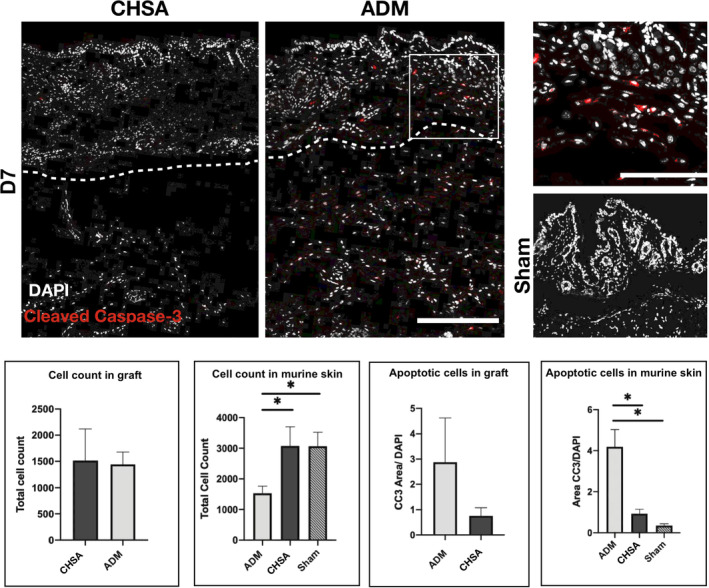
Figure 2.

Immunofluorescent staining for cleaved caspase‐3 (CC3) indicating apoptotic cells after implantation of cryopreserved human skin allografts (CHSA), acellular dermal matrix (ADM), or sham surgeries. White box indicating the area of the magnified image on the right. Dotted lines indicate the interface between murine dermis and xenografts. The white square indicates the area of the magnified image. Scale bar: 200 μm in overview and 100 μm in magnified image.*P < .05
