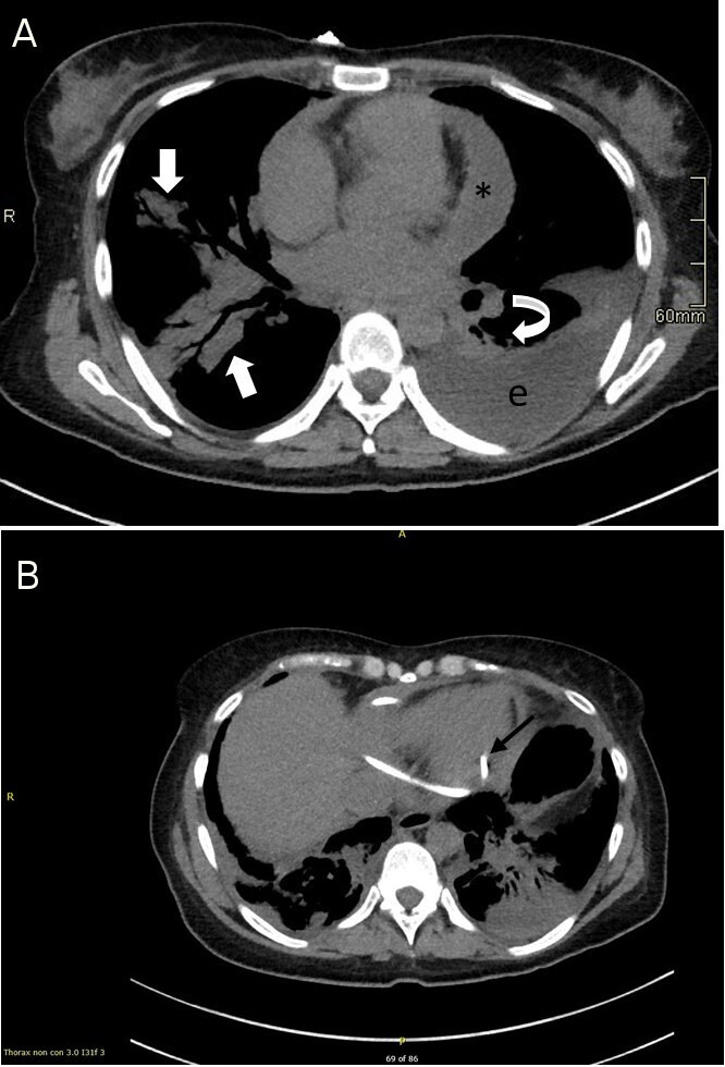
Figure 3.

(A) CT of thorax: Large pericardial effusion (asterisk (*)). Large left-sided loculated pleural effusion (e) with overlying consolidation in the left lower lobe (curved block arrow). Small right-sided pleural effusion, with patchy peribronchial consolidation (block arrows) involving the right middle and lower lobes. (B) CT of thorax: Image demonstrates pericardial drain in situ with the tip (arrow) sited postero-inferior to the left ventricle.
