
Figure 5.
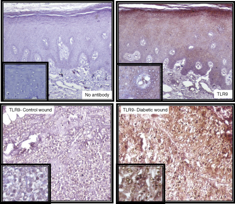
Immunohistochemistry of TLR9 in wound samples (3‐µm‐thick dewaxed paraffin sections) with insets showing detail of staining (40× magnifications). Upper panel shows the negative (left) and positive (right) staining. In the negative control, serum and secondary antibodies were applied but no primary TLR9 antibody was added to the staining solution to check the non‐specific binding of the primary antibody. Lower panel shows the immunohistochemistry for TLR9 in non‐diabetic control wound (left) and diabetic wound (right).